Difference between revisions of "ADDUCTOR CANAL"
(Imported from text file) |
(Imported from text file) |
||
| Line 1: | Line 1: | ||
[[Summary Article| | ===== [[Summary Article|'''SUMMARY''']] ===== | ||
1. Also known as the Hunter's canal. | |||
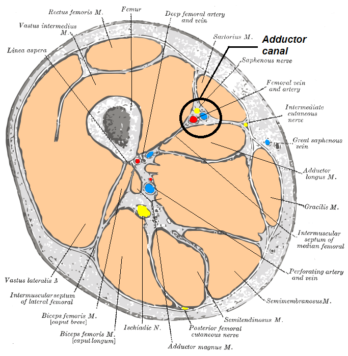
<br/> 2. Formed in the gutter b/w the vastus medialis & adductor longus (superiorly) and vastus medialis & adductor magnus (inferiorly). The sartorius forms its roof. | <br/> 2. Formed in the gutter b/w the vastus medialis & adductor longus (superiorly) and vastus medialis & adductor magnus (inferiorly). The sartorius forms its roof. | ||
<br/>3. Continues from the apex of the femoral triangle. | <br/>3. Continues from the apex of the femoral triangle. | ||
<br/>4. Roofed by fascia which contains the subsartorial plexus. | |||
<br/> | <br/> | ||
<br/>5. Contents - femoral artery, vein, saphenous nerve. In the superior part - nerve to the vastus medialis. | <br/>5. Contents - femoral artery, vein, saphenous nerve. In the superior part - nerve to the vastus medialis. | ||
<br/>[[Image:Adductor_canal.png]] | <br/>[[Image:Adductor_canal.png]] | ||
<br/> | <br/> | ||
<br/><b>Image:</b> Häggström, Mikael (2014). <i>Medical gallery of Mikael Häggström 2014.</i> WikiJournal of Medicine 1 (2). DOI:10.15347/wjm/2014.008. ISSN 2002-4436. [Public domain], [https://commons.wikimedia.org/wiki/File:Adductor_canal.png via Wikimedia Commons] [Accessed 20 Apr. 2019]. | <br/><b>Image:</b> Häggström, Mikael (2014). <i>Medical gallery of Mikael Häggström 2014.</i> WikiJournal of Medicine 1 (2). DOI:10.15347/wjm/2014.008. ISSN 2002-4436. [Public domain], [https://commons.wikimedia.org/wiki/File:Adductor_canal.png via Wikimedia Commons] [Accessed 20 Apr. 2019]. | ||
==Reference(s)== | ==Reference(s)== | ||
Revision as of 08:38, 30 December 2022
SUMMARY
1. Also known as the Hunter's canal.
2. Formed in the gutter b/w the vastus medialis & adductor longus (superiorly) and vastus medialis & adductor magnus (inferiorly). The sartorius forms its roof.
3. Continues from the apex of the femoral triangle.
4. Roofed by fascia which contains the subsartorial plexus.
5. Contents - femoral artery, vein, saphenous nerve. In the superior part - nerve to the vastus medialis.

Image: Häggström, Mikael (2014). Medical gallery of Mikael Häggström 2014. WikiJournal of Medicine 1 (2). DOI:10.15347/wjm/2014.008. ISSN 2002-4436. [Public domain], via Wikimedia Commons [Accessed 20 Apr. 2019].
Reference(s)
R.M.H McMinn (1998). Last’s anatomy: regional and applied. Edinburgh: Churchill Livingstone.
Gray, H., Carter, H.V. and Davidson, G. (2017). Gray’s anatomy. London: Arcturus.