Difference between revisions of "OBTURATOR NERVE"
(Imported from text file) |
|||
| (2 intermediate revisions by the same user not shown) | |||
| Line 1: | Line 1: | ||
[[Summary Article| | ===== [[Summary Article|'''SUMMARY''']] ===== | ||
SUMMARY | |||
<br/>1. Origin - L2 + 3 + 4. | <br/>1. Origin - L2 + 3 + 4. | ||
<br/>2. Arises from the medial side of the psoas, lies on the sacral ala, lateral to the lumbosacral trunk. | <br/>2. Arises from the medial side of the psoas, lies on the sacral ala, lateral to the lumbosacral trunk. | ||
<br/>3. From the angle of the external & internal iliac vessels it runs to the obturator foramen. In the obturator canal it divides into POSTERIOR and ANTERIOR divisons (which lie posterior and anterior to the ADDUCTOR BREVIS respectively). | <br/>3. From the angle of the external & internal iliac vessels it runs to the obturator foramen. In the obturator canal it divides into POSTERIOR and ANTERIOR divisons (which lie posterior and anterior to the ADDUCTOR BREVIS respectively). | ||
<br/> | <br/> | ||
<br/> | <br/>SUPPLY | ||
<br/>4. MAIN NERVE - supplies the parietal peritoneum of the side wall of the pelvis. | <br/>4. MAIN NERVE - supplies the parietal peritoneum of the side wall of the pelvis. | ||
<br/>5. POSTERIOR DIVISION - supplies the obturator externus, adductor magnus (with the sciatic nerve), knee joint. | <br/>5. POSTERIOR DIVISION - supplies the obturator externus, adductor magnus (with the sciatic nerve), knee joint. | ||
<br/>6. ANTERIOR DIVISION - supplies the adductor brevis, adductor longus, pectineus (with the femoral nerve), gracilis. | <br/>6. ANTERIOR DIVISION - supplies the adductor brevis, adductor longus, pectineus (with the femoral nerve), gracilis. | ||
<br/>7. CUTANEOUS BRANCH OF ANTERIOR DIVISION - supplies the lower medial side of thigh. | <br/>7. CUTANEOUS BRANCH OF ANTERIOR DIVISION - supplies the lower medial side of thigh. | ||
<br/>[[Image:Gray837.png]] | <br/>[[Image:Gray837.png]] | ||
<br/><b>Image: | <br/> | ||
<br/><b>Image: </b>Henry Vandyke Carter [Public domain], [https://commons.wikimedia.org/wiki/File:Gray837.png via Wikimedia Commons] [Accessed 28 Sep. 2019]. | |||
==Reference(s)== | ==Reference(s)== | ||
Latest revision as of 11:29, 1 January 2023
SUMMARY
SUMMARY
1. Origin - L2 + 3 + 4.
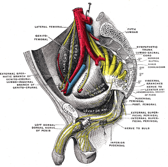
2. Arises from the medial side of the psoas, lies on the sacral ala, lateral to the lumbosacral trunk.
3. From the angle of the external & internal iliac vessels it runs to the obturator foramen. In the obturator canal it divides into POSTERIOR and ANTERIOR divisons (which lie posterior and anterior to the ADDUCTOR BREVIS respectively).
SUPPLY
4. MAIN NERVE - supplies the parietal peritoneum of the side wall of the pelvis.
5. POSTERIOR DIVISION - supplies the obturator externus, adductor magnus (with the sciatic nerve), knee joint.
6. ANTERIOR DIVISION - supplies the adductor brevis, adductor longus, pectineus (with the femoral nerve), gracilis.
7. CUTANEOUS BRANCH OF ANTERIOR DIVISION - supplies the lower medial side of thigh.

Image: Henry Vandyke Carter [Public domain], via Wikimedia Commons [Accessed 28 Sep. 2019].
Reference(s)
R.M.H McMinn (1998). Last’s anatomy: regional and applied. Edinburgh: Churchill Livingstone.
Gray, H., Carter, H.V. and Davidson, G. (2017). Gray’s anatomy. London: Arcturus.