Difference between revisions of "GREAT SAPHENOUS VEIN"
(Imported from text file) |
(Imported from text file) |
||
| (2 intermediate revisions by the same user not shown) | |||
| Line 1: | Line 1: | ||
[[Summary Article| | ===== [[Summary Article|'''SUMMARY''']] ===== | ||
1. Longest vein in the body. | |||
<br/> | <br/> | ||
<br/>2. Continuation of the medial marginal vein of the foot. | <br/>2. Continuation of the medial marginal vein of the foot. | ||
<br/> | <br/> | ||
<br/>3. Courses in front of the medial malleolus = | <br/>3. Courses in front of the medial malleolus => crosses the medial surface of the tibia obliquely towards the knee => where it lies a hands-breath behind the medial border of the patella. | ||
<br/> | <br/> | ||
<br/> | <br/>4. It spirals forwards around the medial convexity of the thigh and ends by passing through the cribriform fascia covering the saphenous opening (3.5cm below and lateral to the pubic tubercle). | ||
<br/> | <br/> | ||
<br/>6. It may also be joined by the anterolateral and posteromedial femoral veins here. | <br/>5. Here it joins the femoral vein along with 4 others (superficial circumflex iliac, superficial epigastric, superficial and deep external peudendal veins). | ||
<br/> | |||
<br/>6. It may also be joined by the anterolateral and posteromedial femoral veins here. | |||
<br/> | <br/> | ||
<br/>[[Image:Great_saphenous_vein.png]] | <br/>[[Image:Great_saphenous_vein.png]] | ||
<br/> | <br/> | ||
<br/><b>Image:</b> | <br/><b>Image:</b> Häggström, Mikael (2014). <i>Medical gallery of Mikael Häggström 2014.</i> WikiJournal of Medicine 1 (2). DOI:10.15347/wjm/2014.008. ISSN 2002-4436. [Public domain], [https://commons.wikimedia.org/wiki/File:Adductor_canal.png via Wikimedia Commons] [Accessed 24 Sep. 2019]. | ||
==Reference(s)== | ==Reference(s)== | ||
Latest revision as of 11:29, 1 January 2023
SUMMARY
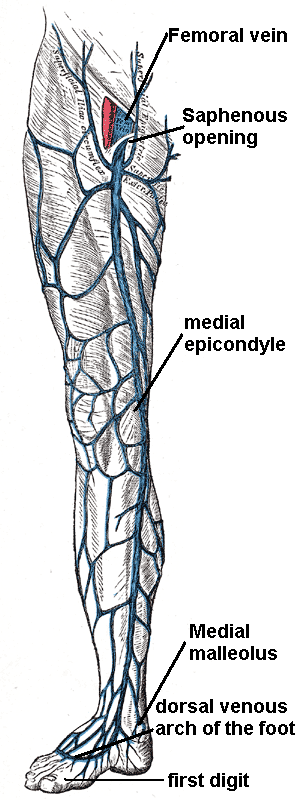
1. Longest vein in the body.
2. Continuation of the medial marginal vein of the foot.
3. Courses in front of the medial malleolus => crosses the medial surface of the tibia obliquely towards the knee => where it lies a hands-breath behind the medial border of the patella.
4. It spirals forwards around the medial convexity of the thigh and ends by passing through the cribriform fascia covering the saphenous opening (3.5cm below and lateral to the pubic tubercle).
5. Here it joins the femoral vein along with 4 others (superficial circumflex iliac, superficial epigastric, superficial and deep external peudendal veins).
6. It may also be joined by the anterolateral and posteromedial femoral veins here.

Image: Häggström, Mikael (2014). Medical gallery of Mikael Häggström 2014. WikiJournal of Medicine 1 (2). DOI:10.15347/wjm/2014.008. ISSN 2002-4436. [Public domain], via Wikimedia Commons [Accessed 24 Sep. 2019].
Reference(s)
R.M.H McMinn (1998). Last’s anatomy: regional and applied. Edinburgh: Churchill Livingstone.
Gray, H., Carter, H.V. and Davidson, G. (2017). Gray’s anatomy. London: Arcturus.