Difference between revisions of "FEMORAL ARTERY"
(Imported from text file) |
(Imported from text file) |
||
| (2 intermediate revisions by the same user not shown) | |||
| Line 1: | Line 1: | ||
[[Summary Article| | ===== [[Summary Article|'''SUMMARY''']] ===== | ||
1. Enters the thigh midway b/w the ASIS and pubic symphysis, just medial to the deep inguinal ring. | |||
<br/>2. Here it lies on the psoas major which separates it from the capsule of the hip joint. | <br/>2. Here it lies on the psoas major which separates it from the capsule of the hip joint. | ||
<br/>3. It emerges from the femoral sheath to disappear beneath the sartorius into the adductor canal. | <br/>3. It emerges from the femoral sheath to disappear beneath the sartorius into the adductor canal. | ||
<br/>4. Gives off profunda femoris & 4 small branches. | |||
<br/> | <br/> | ||
<br/>5. Surface marking - from the midpoint b/w ASIS & pubic symphysis, along the upper 2/3 of a line to the adductor tubercle. | |||
<br/>5. Surface marking - from the midpoint b/w ASIS & pubic symphysis, along the upper 2/3 of a line to the adductor tubercle. | <br/><i>6. At all levels in the thigh it lies b/w the saphenous nerve and femoral vein. </i> | ||
<br/><i>6. At all levels in the thigh it lies b/w the saphenous nerve and femoral vein. | |||
<br/><i>[[Image:Sobo_1909_573-574.png]]</i> | <br/><i>[[Image:Sobo_1909_573-574.png]]</i> | ||
<br/><b>Image:</b> | <br/><b>Image:</b> Dr. Johannes Sobotta [Public domain], [https://commons.wikimedia.org/wiki/File:Sobo_1909_573-574.png via Wikimedia Commons] [Accessed 7 Apr. 2019]. | ||
==Reference(s)== | ==Reference(s)== | ||
Latest revision as of 11:29, 1 January 2023
SUMMARY
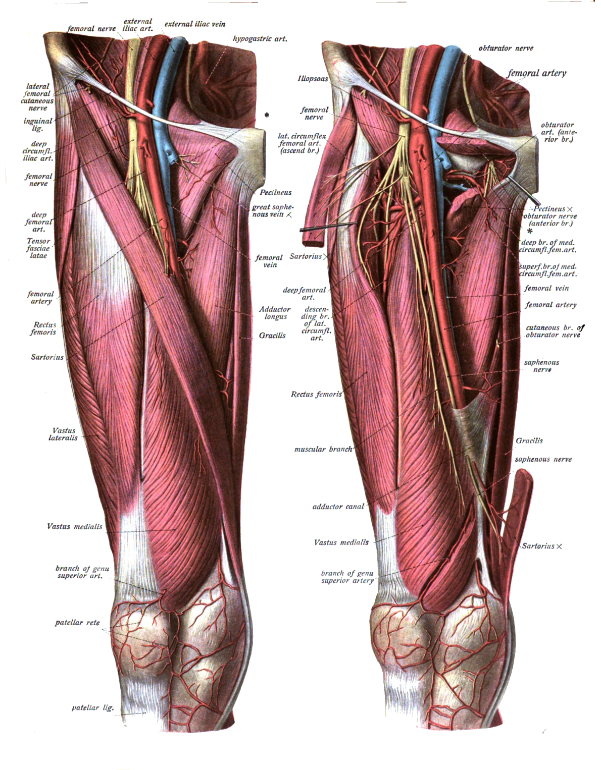
1. Enters the thigh midway b/w the ASIS and pubic symphysis, just medial to the deep inguinal ring.
2. Here it lies on the psoas major which separates it from the capsule of the hip joint.
3. It emerges from the femoral sheath to disappear beneath the sartorius into the adductor canal.
4. Gives off profunda femoris & 4 small branches.
5. Surface marking - from the midpoint b/w ASIS & pubic symphysis, along the upper 2/3 of a line to the adductor tubercle.
6. At all levels in the thigh it lies b/w the saphenous nerve and femoral vein.

Image: Dr. Johannes Sobotta [Public domain], via Wikimedia Commons [Accessed 7 Apr. 2019].
Reference(s)
R.M.H McMinn (1998). Last’s anatomy: regional and applied. Edinburgh: Churchill Livingstone.
Gray, H., Carter, H.V. and Davidson, G. (2017). Gray’s anatomy. London: Arcturus.