Difference between revisions of "THORACIC VERTEBRA-COSTAL FACETS"
(Imported from text file) |
|||
| Line 11: | Line 11: | ||
<br/>[[Image:bones-and-ligaments-of-the-vertebral-column-illustrations-14287c875e0410c23295ad6ce323663f0e92b5a1.jpg]] | <br/>[[Image:bones-and-ligaments-of-the-vertebral-column-illustrations-14287c875e0410c23295ad6ce323663f0e92b5a1.jpg]] | ||
<br/> | <br/> | ||
<br/><b>Image:</b> Case courtesy of OpenStax College, [https://radiopaedia.org/?lang=gb Radiopaedia.org]. From the case [https://radiopaedia.org/cases/42770?lang=gb rID: 42770]. | <br/><b>Image: </b>Case courtesy of OpenStax College, [https://radiopaedia.org/?lang=gb Radiopaedia.org]. From the case [https://radiopaedia.org/cases/42770?lang=gb rID: 42770]. | ||
Latest revision as of 12:32, 27 March 2023
SUMMARY
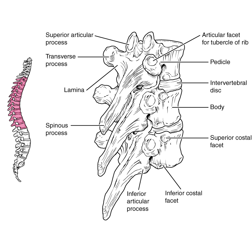
1. The costal facets differentiate thoracic vertebrae from others
2. Part of the morphological neural arch and not the centrum
3. Actually consists of 2 pairs of demifacets, each (covered by hyaline cartilage) makes a separate synovial joint with the facet of the rib head
4. The upper demifacet lies between the upper border of the body & upper border of the pedicle, semicircular in outline & lies vertical
5. The lower demifacet lies on the lower border of the body, smaller & faces downwards

Image: Case courtesy of OpenStax College, Radiopaedia.org. From the case rID: 42770.
Reference(s)
R.M.H McMinn (1998). Last’s anatomy: regional and applied. Edinburgh: Churchill Livingstone. Get it on Amazon.
Drake, Richard L., et al. Gray's Anatomy for Students. Elsevier, 2023. Get it on Amazon.