Difference between revisions of "CARDIAC ELECTRICAL ACTIVITY-NODES"
From NeuroRehab.wiki
(Imported from text file) |
(Imported from text file) |
||
| Line 14: | Line 14: | ||
==Reference(s)== | ==Reference(s)== | ||
Barrett, K.E., Barman, S.M | Barrett, K.E., Barman, S.M., Brooks, H.L., X, J. and Ganong, W.F. (2019). Ganong’s review of medical physiology. 26th ed. New York: Mcgraw-Hill Education | ||
[[Category:Cardiac Electrical Activity]] | [[Category:Cardiac Electrical Activity]] | ||
[[Category:Physiology]] | [[Category:Physiology]] | ||
Latest revision as of 02:30, 21 March 2023
SUMMARY
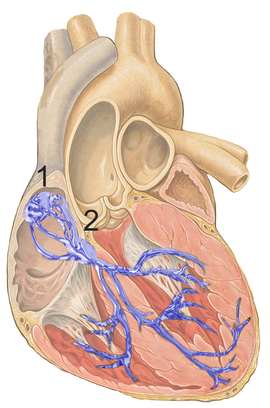
1. Conducts impulses from SA node ⟹ AV node ⟹ bundle of His ⟹ right & left branches ⟹ subendocardial Purkinje fibers.
2. SA node: situated in the right atrium, just below the SVC, atop the crista terminalis. Contains P (pacemaker) cells.
3. AV node: situated in the IA septum, above & to the left of opening of coronary sinus.
4. Because the fibrous framework of the heart separates the atria from ventricles, the bundle is the only means of conducting normal impulses from the atria to ventricles.

Image: J. Heuser, CC BY 2.5 [Accessed 7 Apr. 2019].
Reference(s)
Barrett, K.E., Barman, S.M., Brooks, H.L., X, J. and Ganong, W.F. (2019). Ganong’s review of medical physiology. 26th ed. New York: Mcgraw-Hill Education