Difference between revisions of "RADIAL ARTERY-FOREARM"
(Imported from text file) |
(Imported from text file) |
||
| (2 intermediate revisions by the same user not shown) | |||
| Line 1: | Line 1: | ||
[[Summary Article| | ===== [[Summary Article|'''SUMMARY''']] ===== | ||
1. Arises from the bifurcation of the brachial artery in the cubital fossa. | |||
<br/> | <br/> | ||
<br/>3. It then passes over the | <br/>2. The radial artery passes medial to the biceps tendon, overlapped by the brachioradialis. | ||
<br/> | |||
<br/>3. It then passes over the supinator ⟹ pronator teres ⟹ FDS ⟹ FPL ⟹ PQ ⟹ radius. | |||
<br/> | |||
<br/>4. The radial nerve lies lateral to the middle 1/3 of the artery. | <br/>4. The radial nerve lies lateral to the middle 1/3 of the artery. | ||
<br/>5. It disappears beneath the tendons of the APB & EPL to cross the anatomical snuff box. | <br/> | ||
<br/><i>6. Branch | <br/>5. It disappears beneath the tendons of the APB & EPL to cross the anatomical snuff box. | ||
<br/><i>7. Surface marking | <br/> | ||
<br/><i>6. Branch: superficial palmar branch (completes the superficial palmar arch with the ulnar a.)</i> | |||
<br/> | |||
<br/><i>7. Surface marking: along a line medial to the biceps tendon in the cubital fossa to a point medial to the styloid process.</i> | |||
<br/> | <br/> | ||
<br/>[[Image:Gray528.png]] | <br/>[[Image:Gray528.png]] | ||
<br/><b>Image: | <br/> | ||
<br/><b>Image: </b>Gray, Henry. <i>Anatomy of the Human Body.</i> Philadelphia: Lea & Febiger, 1918; Bartleby.com, 2000. [https://www.bartleby.com/107/ www.bartleby.com/107/] [Accessed 17 Apr. 2019]. | |||
==Reference(s)== | ==Reference(s)== | ||
Latest revision as of 18:52, 8 January 2023
SUMMARY
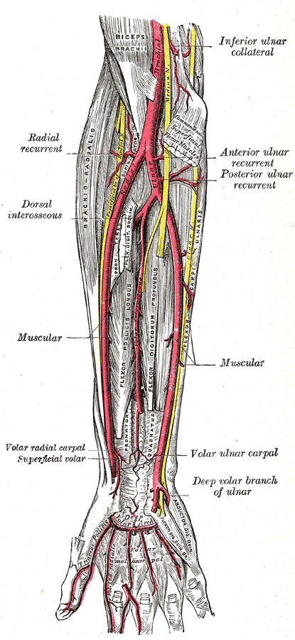
1. Arises from the bifurcation of the brachial artery in the cubital fossa.
2. The radial artery passes medial to the biceps tendon, overlapped by the brachioradialis.
3. It then passes over the supinator ⟹ pronator teres ⟹ FDS ⟹ FPL ⟹ PQ ⟹ radius.
4. The radial nerve lies lateral to the middle 1/3 of the artery.
5. It disappears beneath the tendons of the APB & EPL to cross the anatomical snuff box.
6. Branch: superficial palmar branch (completes the superficial palmar arch with the ulnar a.)
7. Surface marking: along a line medial to the biceps tendon in the cubital fossa to a point medial to the styloid process.

Image: Gray, Henry. Anatomy of the Human Body. Philadelphia: Lea & Febiger, 1918; Bartleby.com, 2000. www.bartleby.com/107/ [Accessed 17 Apr. 2019].
Reference(s)
R.M.H McMinn (1998). Last’s anatomy: regional and applied. Edinburgh: Churchill Livingstone.
Gray, H., Carter, H.V. and Davidson, G. (2017). Gray’s anatomy. London: Arcturus.