Difference between revisions of "MRI-TERMINOLOGY"
(Imported from text file) |
|
| (One intermediate revision by one other user not shown) | |
(No difference)
| |
Latest revision as of 09:55, 25 July 2023
SUMMARY
When describing most MRI sequences we refer to the shade of grey of tissues or fluid with the word intensity, leading to the following absolute terms:
1. High signal intensity ⟹ white
2. Intermediate signal intensity ⟹ grey
3. Low signal intensity ⟹ black
Often we refer to the appearance by relative terms:
1. Hyperintense ⟹ brighter than the thing we are comparing it to
2. Isointense ⟹ same brightness as the thing we are comparing it to
3. Hypointense ⟹ darker than the thing we are comparing it to

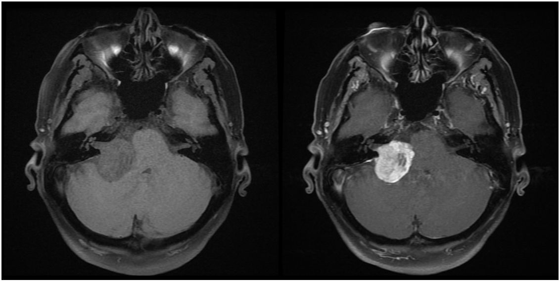
Images: The above images shows a lesion that is isointense on T1W on the left, and hyperintense on T1W with gadolinium on the right.
Reference(s)
Furman, Michael B., and Leland Berkwits. Atlas of Image-Guided Spinal Procedures. Elsevier, Inc, 2017.
Horowitz AL. MRI Physics for Physicians. Springer Science & Business Media. (1989) ISBN:1468403338.
Mangrum W, Christianson K, Duncan S et-al. Duke Review of MRI Principles. Mosby. (2012) ISBN:1455700843.