Difference between revisions of "THORACIC VERTEBRA"
(Imported from text file) |
(Imported from text file) |
||
| (One intermediate revision by one other user not shown) | |||
| Line 13: | Line 13: | ||
<br/>[[Image:bones-and-ligaments-of-the-vertebral-column-illustrations-14287c875e0410c23295ad6ce323663f0e92b5a1.jpg]] | <br/>[[Image:bones-and-ligaments-of-the-vertebral-column-illustrations-14287c875e0410c23295ad6ce323663f0e92b5a1.jpg]] | ||
<br/> | <br/> | ||
<br/><b>Image:</b>Case courtesy of OpenStax College, [https://radiopaedia.org/?lang=gb Radiopaedia.org]. From the case [https://radiopaedia.org/cases/42770?lang=gb rID: 42770]. | <br/><b>Image: </b>Case courtesy of OpenStax College, [https://radiopaedia.org/?lang=gb Radiopaedia.org]. From the case [https://radiopaedia.org/cases/42770?lang=gb rID: 42770]. | ||
Latest revision as of 12:32, 27 March 2023
SUMMARY
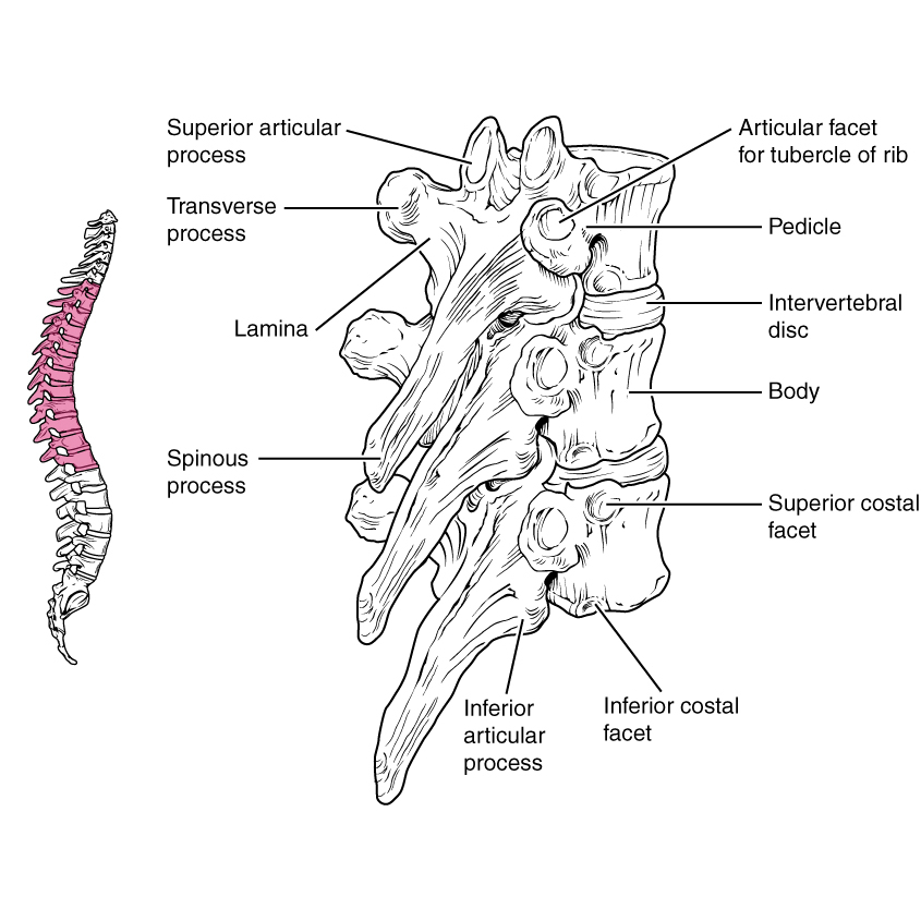
1. Normally kyphotic (hunchback, bent forward)
2. Costal elements ossify separately as ribs
3. The concave facets on the upper transverse processes (TP) look downwards, whilst the flat facets on the lower TP looks upwards
4. The spinous processes (SP) slope downwards from T1-7, from T7-12 become progressively more horizontal
5. The prominent SP of the vertebra prominence may be that of C7 or T1
6. The vertebral bodies become larger from above downwards with the middle 4 showing slight asymmetry with a left-sided excavation produced by the descending aorta

Image: Case courtesy of OpenStax College, Radiopaedia.org. From the case rID: 42770.
Reference(s)
R.M.H McMinn (1998). Last’s anatomy: regional and applied. Edinburgh: Churchill Livingstone. Get it on Amazon.
Drake, Richard L., et al. Gray's Anatomy for Students. Elsevier, 2023. Get it on Amazon.