Difference between revisions of "ULNAR NERVE-E.WRIST & HAND"
(Imported from text file) |
(Imported from text file) |
||
| (One intermediate revision by the same user not shown) | |||
| Line 1: | Line 1: | ||
===== [[Summary Article|'''SUMMARY''']] ===== | ===== [[Summary Article|'''SUMMARY''']] ===== | ||
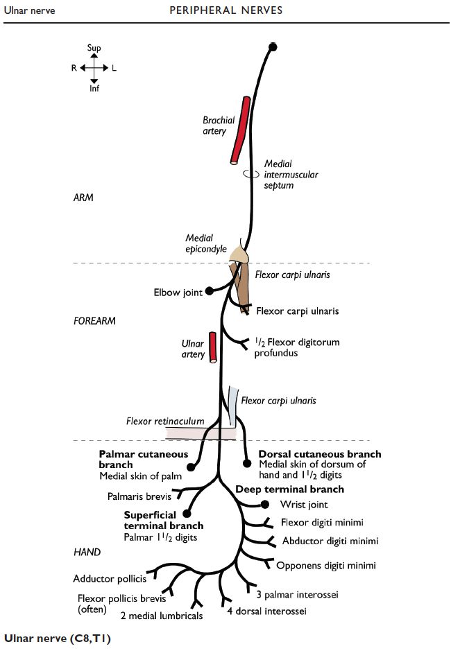
1. The ulnar nerve and artery (lateral to the nerve) enter the hand on the flexor retinaculum, close to the pisiform. Both pass through the Guyon canal. | 1. The ulnar nerve and artery (lateral to the nerve) enter the hand on the flexor retinaculum, close to the pisiform. Both pass through the Guyon canal. | ||
<br/>2. The ulnar nerve divides into the <i>superficial (cutaneous) & deep (muscular) branches</i> at the distal border of the retinaculum. | <br/>2. The ulnar nerve divides into the <i>superficial (cutaneous) & deep (muscular) branches</i> at the distal border of the retinaculum. | ||
<br/> | <br/> | ||
<br/>SUPERFICIAL CUTANEOUS BRANCH | <br/><i>SUPERFICIAL CUTANEOUS BRANCH</i> | ||
<br/><i>3. Supplies palmar skin over medial 1.5 digits. | <br/><i>3. Supplies palmar skin over medial 1.5 digits. </i> | ||
<br/> | <br/> | ||
<br/>DEEP MOTOR BRANCH | <br/><i>DEEP MOTOR BRANCH</i> | ||
<br/><i>4. Supplies medial 2 lumbricals, all interossi, all hypothenar muscles, adductor pollicis (of the thumb). | <br/><i>4. Supplies medial 2 lumbricals, all interossi, all hypothenar muscles, adductor pollicis (of the thumb). </i> | ||
<br/> | <br/> | ||
<br/><i>[[Image:paste-53437983097771.jpg]]</i> | <br/><i>[[Image:paste-53437983097771.jpg]]</i> | ||
<br/> | <br/> | ||
<br/><b>Image:</b> | <br/><b>Image:</b> Whitaker, R. and Borley, N. (2016). Instant anatomy. 6th ed. Chichester (West Sussex): Wiley Blackwell, p.126. | ||
Latest revision as of 18:52, 8 January 2023
SUMMARY
1. The ulnar nerve and artery (lateral to the nerve) enter the hand on the flexor retinaculum, close to the pisiform. Both pass through the Guyon canal.
2. The ulnar nerve divides into the superficial (cutaneous) & deep (muscular) branches at the distal border of the retinaculum.
SUPERFICIAL CUTANEOUS BRANCH
3. Supplies palmar skin over medial 1.5 digits.
DEEP MOTOR BRANCH
4. Supplies medial 2 lumbricals, all interossi, all hypothenar muscles, adductor pollicis (of the thumb).

Image: Whitaker, R. and Borley, N. (2016). Instant anatomy. 6th ed. Chichester (West Sussex): Wiley Blackwell, p.126.
Reference(s)
R.M.H McMinn (1998). Last’s anatomy: regional and applied. Edinburgh: Churchill Livingstone.
Gray, H., Carter, H.V. and Davidson, G. (2017). Gray’s anatomy. London: Arcturus.