Difference between revisions of "ELBOW JOINT-JOINT & CAPSULE"
(Imported from text file) |
(Imported from text file) |
||
| (3 intermediate revisions by the same user not shown) | |||
| Line 1: | Line 1: | ||
[[Summary Article| | ===== [[Summary Article|'''SUMMARY''']] ===== | ||
<i>ARTICULATION</i> | |||
<br/>1. Combination of synovial hinge & pivot joints made up of three articulations. | <br/>1. Combination of synovial hinge & pivot joints made up of three articulations. | ||
<br/> | <br/><i>2. Radiohumeral:</i> capitellum of the humerus with the radial head. | ||
<br/><i>3. Ulnohumeral: </i>trochlea of the humerus with the trochlear notch (with separate olecranon and coronoid process articular facets) of the ulna. | |||
<br/><i>4. Radioulnar:</i> radial head with the radial notch of the ulna (proximal radioulnar joint). | |||
<br/> | <br/> | ||
<br/><i> | <br/><i>FLEXION/EXTENSION</i> | ||
<br/><i>5. In full flexion - </i>the coronoid process is received by the coronoid fossa and the radial head is received by the radial fossa on the anterior surface of the humerus. | <br/><i>5. In full flexion - </i>the coronoid process is received by the coronoid fossa and the radial head is received by the radial fossa on the anterior surface of the humerus. | ||
<br/><i>6. In full extension - </i>the olecranon process is received by the olecranon fossa on the posterior aspect of the humerus. | <br/><i>6. In full extension - </i>the olecranon process is received by the olecranon fossa on the posterior aspect of the humerus. | ||
<br/> | <br/> | ||
<br/>MOVEMENT | <br/><i>MOVEMENT</i> | ||
<br/><i>7. The hinge component</i> (allowing flexion-extension) is formed by the ulnohumeral articulation. | <br/><i>7. The hinge component</i> (allowing flexion-extension) is formed by the ulnohumeral articulation. | ||
<br/><i>8. The pivot component</i> (allowing pronation-supination) is formed by the radiohumeral articulation & proximal radioulnar joints. | <br/><i>8. The pivot component</i> (allowing pronation-supination) is formed by the radiohumeral articulation & proximal radioulnar joints. | ||
<br/> | <br/> | ||
<br/><i>[[Image:Elbow.jpg]]</i> | <br/><i>[[Image:Elbow.jpg]]</i> | ||
<br/><b | <br/> | ||
<br/><b style="font-weight: bold; ">Images: </b>Gray, Henry. <i>Anatomy of the Human Body.</i> Philadelphia: Lea & Febiger, 1918; Bartleby.com, 2000. [https://www.bartleby.com/107/ www.bartleby.com/107/] [Accessed 20 Nov. 2018]. | |||
==Reference(s)== | ==Reference(s)== | ||
Latest revision as of 18:52, 8 January 2023
SUMMARY
ARTICULATION
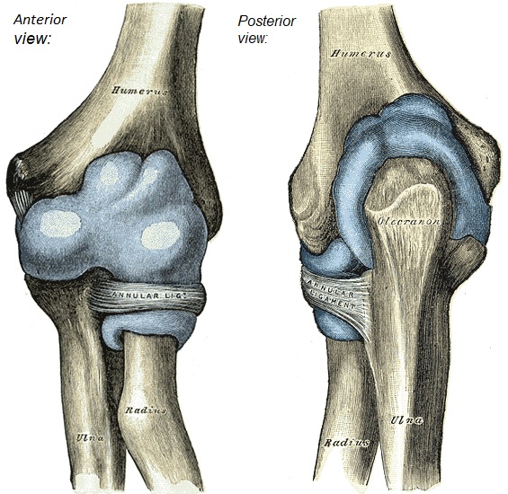
1. Combination of synovial hinge & pivot joints made up of three articulations.
2. Radiohumeral: capitellum of the humerus with the radial head.
3. Ulnohumeral: trochlea of the humerus with the trochlear notch (with separate olecranon and coronoid process articular facets) of the ulna.
4. Radioulnar: radial head with the radial notch of the ulna (proximal radioulnar joint).
FLEXION/EXTENSION
5. In full flexion - the coronoid process is received by the coronoid fossa and the radial head is received by the radial fossa on the anterior surface of the humerus.
6. In full extension - the olecranon process is received by the olecranon fossa on the posterior aspect of the humerus.
MOVEMENT
7. The hinge component (allowing flexion-extension) is formed by the ulnohumeral articulation.
8. The pivot component (allowing pronation-supination) is formed by the radiohumeral articulation & proximal radioulnar joints.

Images: Gray, Henry. Anatomy of the Human Body. Philadelphia: Lea & Febiger, 1918; Bartleby.com, 2000. www.bartleby.com/107/ [Accessed 20 Nov. 2018].
Reference(s)
R.M.H McMinn (1998). Last’s anatomy: regional and applied. Edinburgh: Churchill Livingstone.
Gray, H., Carter, H.V. and Davidson, G. (2017). Gray’s anatomy. London: Arcturus.