Difference between revisions of "MUSCLES-TRICEPS SURAE (SUPERFICIAL POSTERIOR COMPARTMENT)"
(Imported from text file) |
(Imported from text file) |
||
| (2 intermediate revisions by the same user not shown) | |||
| Line 1: | Line 1: | ||
[[Summary Article| | ===== [[Summary Article|'''SUMMARY''']] ===== | ||
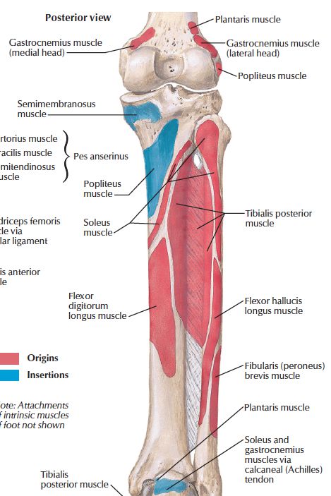
1. GASTROCNEMIUS - <i>lateral head </i>arises from the lateral surface of the lateral femoral condyle. <i>Medial head </i>arises from the medial femoral condyle. The two heads converge with the soleus to form the tendo calcaneus which inserts into the posterior surface of the calcaneus. | |||
<br/> | <br/> | ||
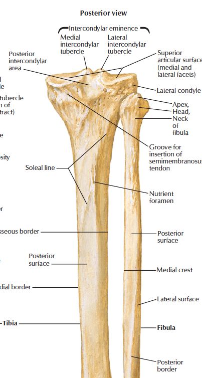
<br/>2. SOLEUS - bipennate muscle that arises from the upper 1/4 of the posterior surface of the fibula + fibrous arch bridging tib/fib + soleal line of the tibia + popliteus fascia above this line. Its combines with the gastrocs to form the tendo calceneus which inserts into the posterior surface of the calcaneus. | <br/>2. SOLEUS - bipennate muscle that arises from the upper 1/4 of the posterior surface of the fibula + fibrous arch bridging tib/fib + soleal line of the tibia + popliteus fascia above this line. Its combines with the gastrocs to form the tendo calceneus which inserts into the posterior surface of the calcaneus. | ||
<br/> | <br/> | ||
<br/>3. PLANTARIS - vestigeal muscle that arises from the upper lateral supracondylar ridge of the femur, lying edge to edge with the lateral head of the gastrocnemius. Its slender tendon runs deep to the medial head of the gastrocnemius to insert into the medial tendo calcaneus. | <br/>3. PLANTARIS - vestigeal muscle that arises from the upper lateral supracondylar ridge of the femur, lying edge to edge with the lateral head of the gastrocnemius. Its slender tendon runs deep to the medial head of the gastrocnemius to insert into the medial tendo calcaneus. | ||
<br/> | <br/> | ||
<br/><i>4. Perforating veins from the great saphenous vein enter the soleus, which contains a rich plexus of veins.</i> | <br/><i>4. Perforating veins from the great saphenous vein enter the soleus, which contains a rich plexus of veins.</i> | ||
<br/ | <br/> | ||
<br/><i>5. All 3 muscles are supplied by the tibial nerve. Aretrial supply - PTA + peroneal a.</i> | <br/><i>5. All 3 muscles are supplied by the tibial nerve. Aretrial supply - PTA + peroneal a.</i> | ||
<br/><i>[[Image:paste-1413044241087.jpg]][[Image:paste-1511828488946.jpg]]</i> | <br/><i>[[Image:paste-1413044241087.jpg]][[Image:paste-1511828488946.jpg]]</i> | ||
==Reference(s)== | ==Reference(s)== | ||
Latest revision as of 11:29, 1 January 2023
SUMMARY
1. GASTROCNEMIUS - lateral head arises from the lateral surface of the lateral femoral condyle. Medial head arises from the medial femoral condyle. The two heads converge with the soleus to form the tendo calcaneus which inserts into the posterior surface of the calcaneus.
2. SOLEUS - bipennate muscle that arises from the upper 1/4 of the posterior surface of the fibula + fibrous arch bridging tib/fib + soleal line of the tibia + popliteus fascia above this line. Its combines with the gastrocs to form the tendo calceneus which inserts into the posterior surface of the calcaneus.
3. PLANTARIS - vestigeal muscle that arises from the upper lateral supracondylar ridge of the femur, lying edge to edge with the lateral head of the gastrocnemius. Its slender tendon runs deep to the medial head of the gastrocnemius to insert into the medial tendo calcaneus.
4. Perforating veins from the great saphenous vein enter the soleus, which contains a rich plexus of veins.
5. All 3 muscles are supplied by the tibial nerve. Aretrial supply - PTA + peroneal a.


Reference(s)
R.M.H McMinn (1998). Last’s anatomy: regional and applied. Edinburgh: Churchill Livingstone.
Gray, H., Carter, H.V. and Davidson, G. (2017). Gray’s anatomy. London: Arcturus.