Difference between revisions of "KNEE JOINT-MENISCI"
(Imported from text file) |
(Imported from text file) |
||
| (2 intermediate revisions by the same user not shown) | |||
| Line 1: | Line 1: | ||
[[Summary Article| | ===== [[Summary Article|'''SUMMARY''']] ===== | ||
1. MEDIAL MENISCUS - longer of the menisci. The anterior horn is attached to the tibial intercondylar area, in front of the ACL. The posterior horn is attached to the tibial intercondylar area, in front of the PCL. <i>It is also attached to the MCL.</i> | |||
<br/ | <br/> | ||
<br/>2. LATERAL MENISCUS - shorter, the anterior and posterior horns are attached immediately in front of & behind the intercondylar spine respectively. The lateral meniscus is attached to the medial femoral condyle via <i>anterior & posterior meniscofemoral ligaments. Its posterior convexity is also attached to the popliteus tendon. | <br/>2. LATERAL MENISCUS - shorter, the anterior and posterior horns are attached immediately in front of & behind the intercondylar spine respectively. The lateral meniscus is attached to the medial femoral condyle via <i>anterior & posterior meniscofemoral ligaments. Its posterior convexity is also attached to the popliteus tendon. </i> | ||
<br/ | <br/> | ||
<br/><i>3. Blood supply - from the peripheral capsule, receive nutrition mostly from the synovial fluid. | <br/><i>3. Blood supply - from the peripheral capsule, receive nutrition mostly from the synovial fluid. </i> | ||
<br/ | <br/> | ||
<br/><i>4. | <br/><i>4. </i><i>The medial mensicus is torn 20x more often than the lateral meniscus due to its relative immobility. </i> | ||
<br/> | <br/> | ||
<br/>[[Image:Gray349.png]] | <br/>[[Image:Gray349.png]] | ||
<br/> | <br/> | ||
<br/><b>Image: | <br/><b>Image: </b>Gray, Henry. <i>Anatomy of the Human Body.</i> Philadelphia: Lea & Febiger, 1918; Bartleby.com, 2000. [https://www.bartleby.com/107/ www.bartleby.com/107/] [Accessed 7 Apr. 2019]. | ||
==Reference(s)== | ==Reference(s)== | ||
Latest revision as of 11:29, 1 January 2023
SUMMARY
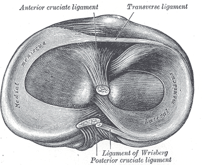
1. MEDIAL MENISCUS - longer of the menisci. The anterior horn is attached to the tibial intercondylar area, in front of the ACL. The posterior horn is attached to the tibial intercondylar area, in front of the PCL. It is also attached to the MCL.
2. LATERAL MENISCUS - shorter, the anterior and posterior horns are attached immediately in front of & behind the intercondylar spine respectively. The lateral meniscus is attached to the medial femoral condyle via anterior & posterior meniscofemoral ligaments. Its posterior convexity is also attached to the popliteus tendon.
3. Blood supply - from the peripheral capsule, receive nutrition mostly from the synovial fluid.
4. The medial mensicus is torn 20x more often than the lateral meniscus due to its relative immobility.

Image: Gray, Henry. Anatomy of the Human Body. Philadelphia: Lea & Febiger, 1918; Bartleby.com, 2000. www.bartleby.com/107/ [Accessed 7 Apr. 2019].
Reference(s)
R.M.H McMinn (1998). Last’s anatomy: regional and applied. Edinburgh: Churchill Livingstone.
Gray, H., Carter, H.V. and Davidson, G. (2017). Gray’s anatomy. London: Arcturus.