Difference between revisions of "HIP JOINT-FEMORAL HEAD & NECK"
(Imported from text file) |
(Imported from text file) |
||
| (One intermediate revision by the same user not shown) | |||
| Line 1: | Line 1: | ||
[[Summary Article| | ===== [[Summary Article|'''SUMMARY''']] ===== | ||
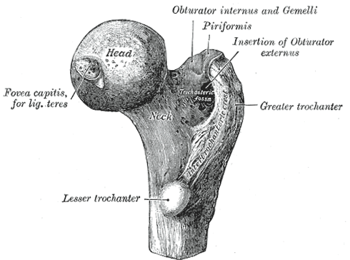
1. The non-articulating convexity of the head is excavated into a pit (the fovea) for attachment of the ligamentum teres. | |||
<br/>2. The other end of the ligamentum teres is attached to the transverse ligament. | <br/>2. The other end of the ligamentum teres is attached to the transverse ligament. | ||
<br/>3. The fibrous capsule is strengthened by 3 ligaments - ilio-femoral, ischio-femoral (<i>weakest ligament)</i>, pubo-femoral. | <br/>3. The fibrous capsule is strengthened by 3 ligaments - ilio-femoral, ischio-femoral (<i>weakest ligament)</i>, pubo-femoral. | ||
<br/>4. These ligaments are 'unwound' if the femur is flexed & externally rotated. | <br/>4. These ligaments are 'unwound' if the femur is flexed & externally rotated. | ||
<br/><i>5. Blood supply - nutrient arteries from the trochanteric anastamosis which travel along the NOF, under the retinacular fibres & artery within the ligamentum teres (usually atrophied by age 7). | <br/><i>5. Blood supply - nutrient arteries from the trochanteric anastamosis which travel along the NOF, under the retinacular fibres & artery within the ligamentum teres (usually atrophied by age 7). </i> | ||
<br/><i>6. Nerve supply - branches of the femoral n., sciatic n. & obturator n.</i> | <br/><i>6. Nerve supply - branches of the femoral n., sciatic n. & obturator n.</i> | ||
<br/ | <br/> | ||
<br/><i>[[Image:Femur_head.png]]</i> | <br/><i>[[Image:Femur_head.png]]</i> | ||
<br/><b>Image: | <br/><b>Image: </b>Gray, Henry. <i>Anatomy of the Human Body.</i> Philadelphia: Lea & Febiger, 1918; Bartleby.com, 2000. [https://www.bartleby.com/107/ www.bartleby.com/107/] [Accessed 14 Apr. 2019]. | ||
==Reference(s)== | ==Reference(s)== | ||
Latest revision as of 11:29, 1 January 2023
SUMMARY
1. The non-articulating convexity of the head is excavated into a pit (the fovea) for attachment of the ligamentum teres.
2. The other end of the ligamentum teres is attached to the transverse ligament.
3. The fibrous capsule is strengthened by 3 ligaments - ilio-femoral, ischio-femoral (weakest ligament), pubo-femoral.
4. These ligaments are 'unwound' if the femur is flexed & externally rotated.
5. Blood supply - nutrient arteries from the trochanteric anastamosis which travel along the NOF, under the retinacular fibres & artery within the ligamentum teres (usually atrophied by age 7).
6. Nerve supply - branches of the femoral n., sciatic n. & obturator n.

Image: Gray, Henry. Anatomy of the Human Body. Philadelphia: Lea & Febiger, 1918; Bartleby.com, 2000. www.bartleby.com/107/ [Accessed 14 Apr. 2019].
Reference(s)
R.M.H McMinn (1998). Last’s anatomy: regional and applied. Edinburgh: Churchill Livingstone.
Gray, H., Carter, H.V. and Davidson, G. (2017). Gray’s anatomy. London: Arcturus.