Difference between revisions of "FEMORAL VEIN"
(Imported from text file) |
(Imported from text file) |
||
| (One intermediate revision by the same user not shown) | |||
| Line 1: | Line 1: | ||
[[Summary Article| | ===== [[Summary Article|'''SUMMARY''']] ===== | ||
1. Continuation of the popliteal vein. | |||
<br/>2. Enters the lower angle of the femoral triangle where it lies posterior to the artery. | <br/>2. Enters the lower angle of the femoral triangle where it lies posterior to the artery. | ||
<br/> | <br/>3. In its course through the triangle it comes to lie medial to the artery in the femoral sheath. | ||
<br/>4. It receives the profunda femoris | <br/>4. It receives the profunda femoris vein & great saphenous vein (just below the femoral sheath, on its anteromedial side). | ||
<br/>5. It passes under the inguinal ligament to run along the pelvic brim as the external iliac vein. | <br/> | ||
<br/>6. It has valves just above the junctions with the profunda femoris and great saphenous veins. | <br/>5. It passes under the inguinal ligament to run along the pelvic brim as the external iliac vein. | ||
<br/>6. It has valves just above the junctions with the profunda femoris and great saphenous veins. | |||
<br/>[[Image:Sobo_1909_573-574.png]] | <br/>[[Image:Sobo_1909_573-574.png]] | ||
<br/><b>Image:</b> | <br/><b>Image:</b> Dr. Johannes Sobotta [Public domain], [https://commons.wikimedia.org/wiki/File:Sobo_1909_573-574.png via Wikimedia Commons] [Accessed 12 Apr. 2019]. | ||
==Reference(s)== | ==Reference(s)== | ||
Latest revision as of 11:29, 1 January 2023
SUMMARY
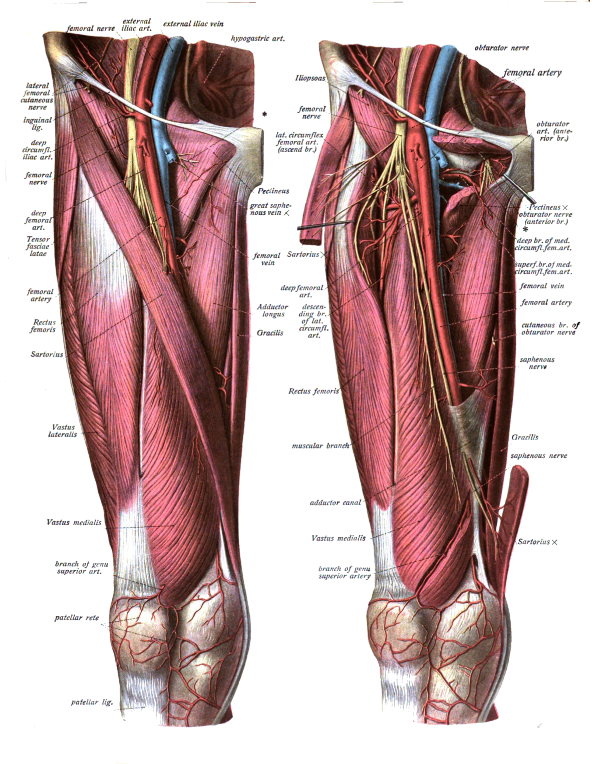
1. Continuation of the popliteal vein.
2. Enters the lower angle of the femoral triangle where it lies posterior to the artery.
3. In its course through the triangle it comes to lie medial to the artery in the femoral sheath.
4. It receives the profunda femoris vein & great saphenous vein (just below the femoral sheath, on its anteromedial side).
5. It passes under the inguinal ligament to run along the pelvic brim as the external iliac vein.
6. It has valves just above the junctions with the profunda femoris and great saphenous veins.

Image: Dr. Johannes Sobotta [Public domain], via Wikimedia Commons [Accessed 12 Apr. 2019].
Reference(s)
R.M.H McMinn (1998). Last’s anatomy: regional and applied. Edinburgh: Churchill Livingstone.
Gray, H., Carter, H.V. and Davidson, G. (2017). Gray’s anatomy. London: Arcturus.