Difference between revisions of "FEMORAL NERVE"
(Imported from text file) |
(Imported from text file) |
||
| (One intermediate revision by the same user not shown) | |||
| Line 1: | Line 1: | ||
[[Summary Article| | ===== [[Summary Article|'''SUMMARY''']] ===== | ||
1. Origin - L2, L3, L4 <i>(posterior divisions of the anterior rami).</i>2. Emerges from the lateral border of the psoas major and runs in the gutter b/w psoas & iliacus, deep to iliac fascia. | |||
<br/>3. Supplies the iliacus, then passes under the inguinal | <br/>3. Supplies the iliacus, then passes under the inguinal ligament & lateral to femoral sheath (remember the mnemonic 'VAN'). | ||
<br/>4. Lateral femoral circumflex artery divides its branches (9-10) into SUPERFICIAL (4 = | <br/>4. Lateral femoral circumflex artery divides its branches (9-10) into SUPERFICIAL (4 => 2 cutaneous + 2 muscular) & DEEP (5-6 => 1 to each vastus [3 total] + 2 to rectus femoris + 1 cutaneous). | ||
<br/>5. SUPERFICIAL BRANCHES - nerve to sartorius, nerve to pectineus, intermediate femoral cutaneous nerve, medial femoral cutaneous nerve. | <br/>5. SUPERFICIAL BRANCHES - nerve to sartorius, nerve to pectineus, intermediate femoral cutaneous nerve, medial femoral cutaneous nerve. | ||
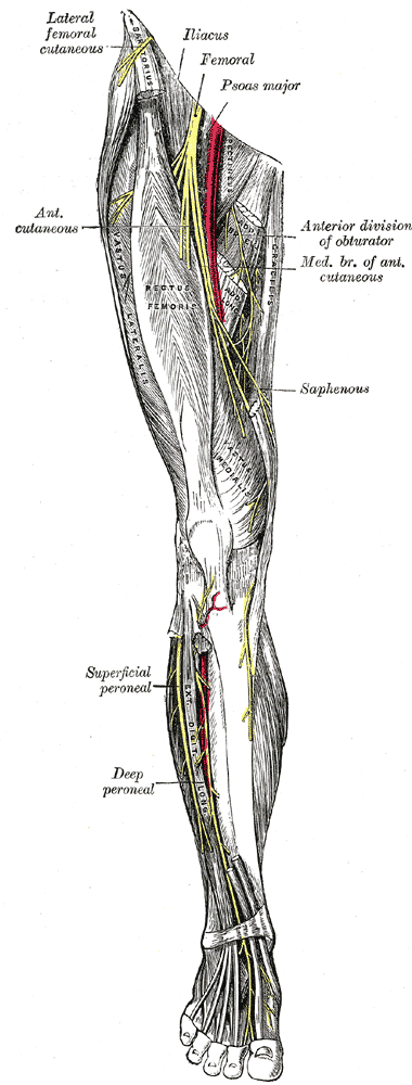
<br/>6. DEEP BRANCHES - 2 nerves to rectus femoris (also supplies hip), nerve to vastus lateralis, nerve to vastus intermedius, nerve to vastus medialis (largest, carries most of branches to knee joint), saphenous nerve. | <br/>6. DEEP BRANCHES - 2 nerves to rectus femoris (also supplies hip), nerve to vastus lateralis, nerve to vastus intermedius, nerve to vastus medialis (largest, carries most of branches to knee joint), saphenous nerve. | ||
<br/>[[Image:Gray827.png]] | <br/>[[Image:Gray827.png]] | ||
<br/><b>Image: | <br/><b>Image: </b>Gray, Henry. <i>Anatomy of the Human Body.</i> Philadelphia: Lea & Febiger, 1918; Bartleby.com, 2000. [https://www.bartleby.com/107/ www.bartleby.com/107/] [Accessed 07 Apr. 2019]. | ||
==Reference(s)== | ==Reference(s)== | ||
Latest revision as of 11:29, 1 January 2023
SUMMARY
1. Origin - L2, L3, L4 (posterior divisions of the anterior rami).2. Emerges from the lateral border of the psoas major and runs in the gutter b/w psoas & iliacus, deep to iliac fascia.
3. Supplies the iliacus, then passes under the inguinal ligament & lateral to femoral sheath (remember the mnemonic 'VAN').
4. Lateral femoral circumflex artery divides its branches (9-10) into SUPERFICIAL (4 => 2 cutaneous + 2 muscular) & DEEP (5-6 => 1 to each vastus [3 total] + 2 to rectus femoris + 1 cutaneous).
5. SUPERFICIAL BRANCHES - nerve to sartorius, nerve to pectineus, intermediate femoral cutaneous nerve, medial femoral cutaneous nerve.
6. DEEP BRANCHES - 2 nerves to rectus femoris (also supplies hip), nerve to vastus lateralis, nerve to vastus intermedius, nerve to vastus medialis (largest, carries most of branches to knee joint), saphenous nerve.

Image: Gray, Henry. Anatomy of the Human Body. Philadelphia: Lea & Febiger, 1918; Bartleby.com, 2000. www.bartleby.com/107/ [Accessed 07 Apr. 2019].
Reference(s)
R.M.H McMinn (1998). Last’s anatomy: regional and applied. Edinburgh: Churchill Livingstone.
Gray, H., Carter, H.V. and Davidson, G. (2017). Gray’s anatomy. London: Arcturus.