Difference between revisions of "QUADRANGULAR SPACE"
From NeuroRehab.wiki
(Imported from text file) |
(Imported from text file) |
||
| Line 1: | Line 1: | ||
===== [[Summary Article|'''SUMMARY''']] ===== | ===== [[Summary Article|'''SUMMARY''']] ===== | ||
1. Lies b/w the subscapularis & teres major in the posterior wall of the axilla. | 1. Lies b/w the subscapularis & teres major in the posterior wall of the axilla. | ||
<br/> | <br/>2. Bounded laterally by the humerus. | ||
<br/>3. Bounded medially by the long head of the triceps. | <br/>3. Bounded medially by the long head of the triceps. | ||
<br/><i>4. Transmits the axillary nerve & posterior circumflex humeral vessels below. | <br/><i>4. Transmits the axillary nerve & posterior circumflex humeral vessels below. </i> | ||
<br/> | |||
<br/><i>[[Image:quadrilateral-space-grays-illustration.jpeg]]</i> | <br/><i>[[Image:quadrilateral-space-grays-illustration.jpeg]]</i> | ||
<br/><b>Image: | <br/><b>Image: </b>Case courtesy of Dr Craig Hacking, [https://radiopaedia.org/ Radiopaedia.org]. From the case [https://radiopaedia.org/cases/67240 rID: 67240] [Accessed 19 Apr. 2019]. | ||
Latest revision as of 11:29, 1 January 2023
SUMMARY
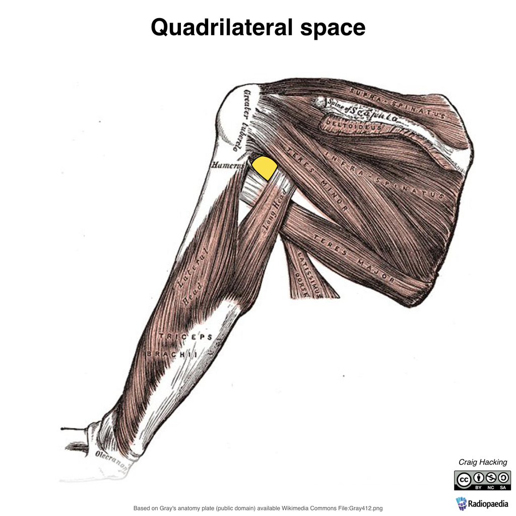
1. Lies b/w the subscapularis & teres major in the posterior wall of the axilla.
2. Bounded laterally by the humerus.
3. Bounded medially by the long head of the triceps.
4. Transmits the axillary nerve & posterior circumflex humeral vessels below.

Image: Case courtesy of Dr Craig Hacking, Radiopaedia.org. From the case rID: 67240 [Accessed 19 Apr. 2019].
Reference(s)
R.M.H McMinn (1998). Last’s anatomy: regional and applied. Edinburgh: Churchill Livingstone.
Gray, H., Carter, H.V. and Davidson, G. (2017). Gray’s anatomy. London: Arcturus.