Difference between revisions of "MUSCLES-ANTERIOR COMPARTMENT OF LEG"
(Imported from text file) |
(Imported from text file) |
||
| Line 1: | Line 1: | ||
[[Summary Article| | ===== [[Summary Article|'''SUMMARY''']] ===== | ||
ALL TENDONS PASS BENEATH THE <b>LATERAL</b> SUPERIOR + INFERIOR EXTENSOR RETINACULUM | |||
<br/> | <br/> | ||
<br/>1. TIBIALIS ANTERIOR - arises from the upper 2/3 of the extensor surface of the tibia + anterior interosseous membrane + overlying deep fascia. Its tendon pierces the superior extensor retinaculum (where it is enclosed by a synovial sheath) + inferior extensor retinaculum to be inserted into the medial cuniform. | <br/>1. TIBIALIS ANTERIOR - arises from the upper 2/3 of the extensor surface of the tibia + anterior interosseous membrane + overlying deep fascia. Its tendon pierces the superior extensor retinaculum (where it is enclosed by a synovial sheath) + inferior extensor retinaculum to be inserted into the medial cuniform. | ||
<br/ | <br/> | ||
<br/>2. EXTENSOR HALLUCIS LONGUS - arises from the middle of the fibula + anterior interosseous membrane. Its tendon passes beneath the superior extensor retinaculum (where it is enclosed by a synovial sheath) + inferior extensor retinaculum to be inserted into the terminal phalanx of the great toe. | <br/>2. EXTENSOR HALLUCIS LONGUS - arises from the middle of the fibula + anterior interosseous membrane. Its tendon passes beneath the superior extensor retinaculum (where it is enclosed by a synovial sheath) + inferior extensor retinaculum to be inserted into the terminal phalanx of the great toe. | ||
<br/ | <br/> | ||
<br/>3. EXTENSOR DIGITORUM LONGUS - arises from the upper 3/4 of the extensor surface of the fibula + tib/fib joint + anterior intermuscular septum + overlying deep fascia. It forms 4 tendons which pass beneath the superior extensor retinaculum + inferior extensor retinaculum (where it shares a synovial sheath with the peroneus tertius tendon) to be inserted into the lateral 4 toes. | <br/>3. EXTENSOR DIGITORUM LONGUS - arises from the upper 3/4 of the extensor surface of the fibula + tib/fib joint + anterior intermuscular septum + overlying deep fascia. It forms 4 tendons which pass beneath the superior extensor retinaculum + inferior extensor retinaculum (where it shares a synovial sheath with the peroneus tertius tendon) to be inserted into the lateral 4 toes. | ||
<br/> | <br/> | ||
| Line 11: | Line 11: | ||
<br/> | <br/> | ||
<br/><i>5. Nerve supply - all are supplied by the deep peroneal nerve. </i> | <br/><i>5. Nerve supply - all are supplied by the deep peroneal nerve. </i> | ||
<br/ | <br/> | ||
<br/><i>6. Arterial supply - branches of the anterior tibial artery. </i> | <br/><i>6. Arterial supply - branches of the anterior tibial artery. </i> | ||
<br/><i>[[Image:paste-4943507358240.jpg]]</i> | <br/><i>[[Image:paste-4943507358240.jpg]]</i> | ||
==Reference(s)== | ==Reference(s)== | ||
Revision as of 08:38, 30 December 2022
SUMMARY
ALL TENDONS PASS BENEATH THE LATERAL SUPERIOR + INFERIOR EXTENSOR RETINACULUM
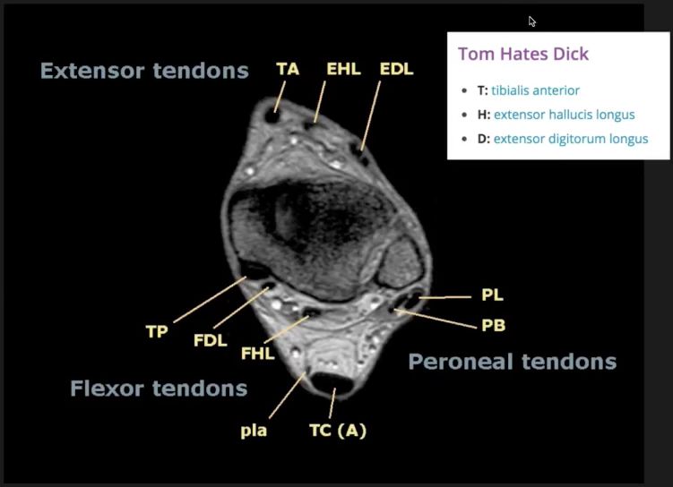
1. TIBIALIS ANTERIOR - arises from the upper 2/3 of the extensor surface of the tibia + anterior interosseous membrane + overlying deep fascia. Its tendon pierces the superior extensor retinaculum (where it is enclosed by a synovial sheath) + inferior extensor retinaculum to be inserted into the medial cuniform.
2. EXTENSOR HALLUCIS LONGUS - arises from the middle of the fibula + anterior interosseous membrane. Its tendon passes beneath the superior extensor retinaculum (where it is enclosed by a synovial sheath) + inferior extensor retinaculum to be inserted into the terminal phalanx of the great toe.
3. EXTENSOR DIGITORUM LONGUS - arises from the upper 3/4 of the extensor surface of the fibula + tib/fib joint + anterior intermuscular septum + overlying deep fascia. It forms 4 tendons which pass beneath the superior extensor retinaculum + inferior extensor retinaculum (where it shares a synovial sheath with the peroneus tertius tendon) to be inserted into the lateral 4 toes.
4. PERONEUS TERTIUS - arises from the lower 1/3 of the fibula beneath the EDL + EHL. Its tendon passes beneath the superior extensor retinaculum + inferior extensor retinaculum (where it shares a synovial sheath with the EDL tendons) to be inserted into the dorsum of the base of the 5th metatarsal.
5. Nerve supply - all are supplied by the deep peroneal nerve.
6. Arterial supply - branches of the anterior tibial artery.

Reference(s)
R.M.H McMinn (1998). Last’s anatomy: regional and applied. Edinburgh: Churchill Livingstone.
Gray, H., Carter, H.V. and Davidson, G. (2017). Gray’s anatomy. London: Arcturus.