Difference between revisions of "DEEP POSTERIOR COMPARTMENT OF FOREARM-EI"
From NeuroRehab.wiki
(Imported from text file) |
(Imported from text file) |
||
| Line 1: | Line 1: | ||
[[Summary Article| | ===== [[Summary Article|'''SUMMARY''']] ===== | ||
EXTENSOR INDICIS | |||
<br/><b><i>TIP: EPL, EI, SUPINATOR arise from the ulna</i></b> | <br/><b><i>TIP: EPL, EI, SUPINATOR arise from the ulna</i></b> | ||
<br/ | <br/> | ||
<br/>1. O: dorsal surface of ulna & interosseous membrane. | <br/>1. O: dorsal surface of ulna & interosseous membrane. | ||
<br/> 2. I: extensor expansion of the index finger. | <br/> 2. I: extensor expansion of the index finger. | ||
| Line 10: | Line 10: | ||
<br/> | <br/> | ||
<br/><b>Image: </b>Extensor indicis muscle. Netter. (2014). Atlas of Human Anatomy, Sixth Edition. 6th ed. Elsevier. | <br/><b>Image: </b>Extensor indicis muscle. Netter. (2014). Atlas of Human Anatomy, Sixth Edition. 6th ed. Elsevier. | ||
==Reference(s)== | ==Reference(s)== | ||
Revision as of 08:38, 30 December 2022
SUMMARY
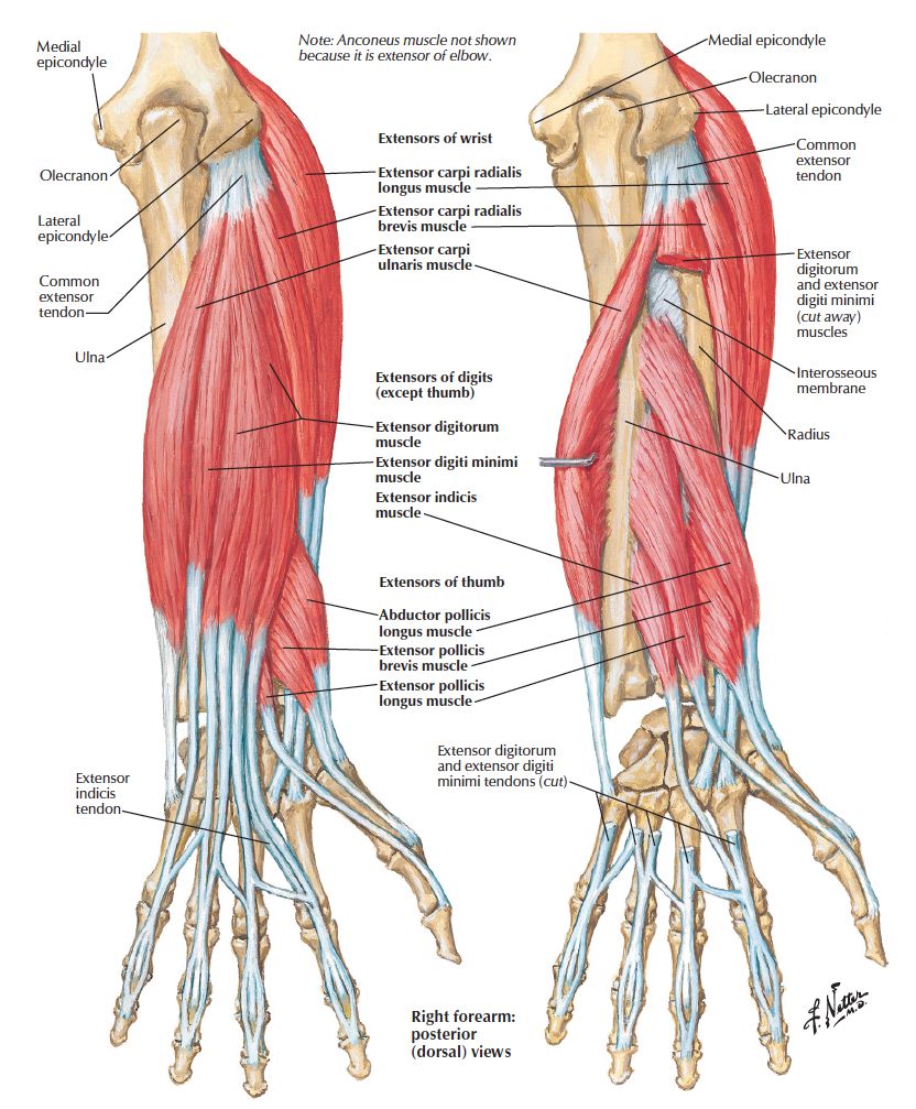
EXTENSOR INDICIS
TIP: EPL, EI, SUPINATOR arise from the ulna
1. O: dorsal surface of ulna & interosseous membrane.
2. I: extensor expansion of the index finger.
3. NS: posterior interosseous branch of radial n.
4. A: extends index finger (2nd digit).

Image: Extensor indicis muscle. Netter. (2014). Atlas of Human Anatomy, Sixth Edition. 6th ed. Elsevier.
Reference(s)
R.M.H McMinn (1998). Last’s anatomy: regional and applied. Edinburgh: Churchill Livingstone.
Gray, H., Carter, H.V. and Davidson, G. (2017). Gray’s anatomy. London: Arcturus.