Difference between revisions of "TROCHANTERIC ANASTAMOSIS"
From NeuroRehab.wiki
(Imported from text file) |
(Imported from text file) |
||
| Line 1: | Line 1: | ||
[[Summary Article|<h5>'''SUMMARY'''</h5>]] | [[Summary Article|<h5>'''SUMMARY'''</h5>]] | ||
<br/>1. Main blood supply of the head of the femur. | <br/>1. Main blood supply of the head of the femur. | ||
<br/> | <br/> 2. Lies near the trochanteric fossa. | ||
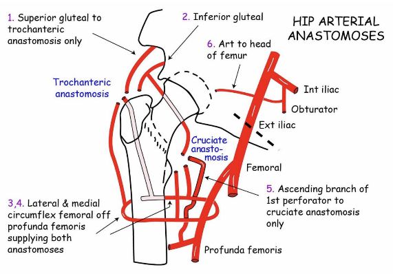
<br/>3. Formed by the anastamosis of the superior, inferior gluteal arteries & medial, lateral circumflex femoral vessels (branches of the profunda femoris artery). | <br/>3. Formed by the anastamosis of the superior, inferior gluteal arteries & medial, lateral circumflex femoral vessels (branches of the profunda femoris artery). | ||
<br/>4. Branches pass along the NOF under the retinacular fibers of the capsule. | <br/>4. Branches pass along the NOF under the retinacular fibers of the capsule. | ||
Revision as of 12:45, 27 December 2022
SUMMARY
1. Main blood supply of the head of the femur.
2. Lies near the trochanteric fossa.
3. Formed by the anastamosis of the superior, inferior gluteal arteries & medial, lateral circumflex femoral vessels (branches of the profunda femoris artery).
4. Branches pass along the NOF under the retinacular fibers of the capsule.

Image: Dr. Appukutty Manickam
Reference(s)
R.M.H McMinn (1998). Last’s anatomy: regional and applied. Edinburgh: Churchill Livingstone.
Gray, H., Carter, H.V. and Davidson, G. (2017). Gray’s anatomy. London: Arcturus.