Difference between revisions of "LATERAL COMPARTMENT OF LEG-PERONEUS LONGUS"
From NeuroRehab.wiki
(Imported from text file) |
(Imported from text file) |
||
| Line 1: | Line 1: | ||
[[Summary Article|<h5>'''SUMMARY'''</h5>]] | [[Summary Article|<h5>'''SUMMARY'''</h5>]] | ||
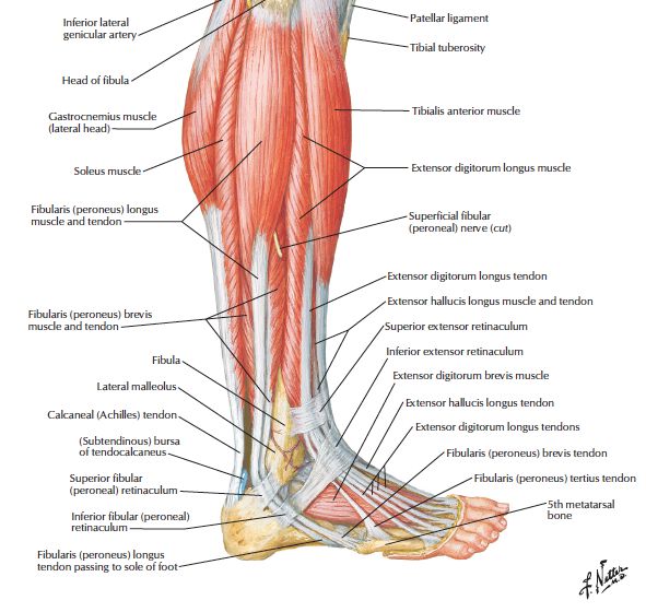
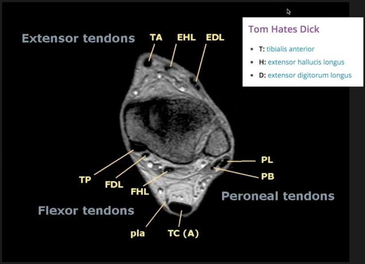
<br/>1. O: head & | <br/>1. O: head & proximal 2/3 of lateral fibula. 2. Its tendon grooves under the LATERAL malleolus (bound down by the superior peroneal retinaculum) & peroneal trochlea of the calcaneus (bound down by the inferior peroneal retinaculum).3. I: crosses the sole of the foot to insert into the 1st MT base & medial cuniform. | ||
<br/> | <br/> | ||
<br/>4. NS: superficial peroneal n. | <br/>4. NS: superficial peroneal n. | ||
<br/>5. A: eversion & plantar-flexion of the foot. | <br/>5. A: eversion & plantar-flexion of the foot. | ||
Revision as of 12:45, 27 December 2022
SUMMARY
1. O: head & proximal 2/3 of lateral fibula. 2. Its tendon grooves under the LATERAL malleolus (bound down by the superior peroneal retinaculum) & peroneal trochlea of the calcaneus (bound down by the inferior peroneal retinaculum).3. I: crosses the sole of the foot to insert into the 1st MT base & medial cuniform.
4. NS: superficial peroneal n.
5. A: eversion & plantar-flexion of the foot.


Image: Netter, F. (2015). Atlas of Human Anatomy. 6th ed. Saint Louis: Elsevier Health Sciences, p.506.
Reference(s)
R.M.H McMinn (1998). Last’s anatomy: regional and applied. Edinburgh: Churchill Livingstone.
Gray, H., Carter, H.V. and Davidson, G. (2017). Gray’s anatomy. London: Arcturus.