Difference between revisions of "OSTEOLOGY-SCAPULA ATTACHMENTS"
(Imported from text file) |
(Imported from text file) |
||
| Line 1: | Line 1: | ||
[[Summary Article|<h5>'''SUMMARY'''</h5>]] | [[Summary Article|<h5>'''SUMMARY'''</h5>]] | ||
<br/>COSTAL SURFACE | |||
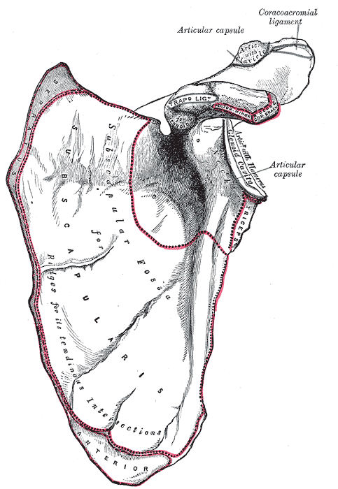
<br/>1. Costal surface/subscapularis fossa - concave, for attachment of the SUBSCAPULARIS. The subscapular bursa separates the lateral bare bone from this muscle. The <i>medial margin of the costal surface </i>receives the SERRATUS ANTERIOR. | <br/>1. Costal surface/subscapularis fossa - concave, for attachment of the SUBSCAPULARIS. The subscapular bursa separates the lateral bare bone from this muscle. The <i>medial margin of the costal surface </i>receives the SERRATUS ANTERIOR. | ||
<br/>SUPERIOR | <br/>SUPERIOR | ||
<br/>2. Suprascapular notch - along the upper border of the scapula, for passage of the <i>suprascapular nerve</i>. The notch is bridged by the transverse scapular ligament. | <br/>2. Suprascapular notch - along the upper border of the scapula, for passage of the <i>suprascapular nerve</i>. The notch is bridged by the transverse scapular ligament. | ||
Revision as of 12:45, 27 December 2022
SUMMARY
COSTAL SURFACE
1. Costal surface/subscapularis fossa - concave, for attachment of the SUBSCAPULARIS. The subscapular bursa separates the lateral bare bone from this muscle. The medial margin of the costal surface receives the SERRATUS ANTERIOR.
SUPERIOR
2. Suprascapular notch - along the upper border of the scapula, for passage of the suprascapular nerve. The notch is bridged by the transverse scapular ligament.
3. Spinoglenoid notch - b/w the scapular spine & glenoid, for passage of the suprascapular n. Common site of impingement.
MEDIAL
4. Medial (vertebral) border - for attachment of the LEVATOR SCAPULAE, RHOMBOID MINOR, RHOMBOID MAJOR.
LATERAL
5. Supraglenoid tubercle - lies on the base of the coracoid, for attachment of the LONG HEAD OF THE BICEPS.
6. Infraglenoid tubercle - along the lateral (axillary) border, for attachment of the LONG HEAD OF THE TRICEPS.
DORSAL
7. Dorsal surface - divided into the supraspinous & infraspinous fossa by the scapular spine, attachment of SUPRASPINATUS & INFRASPINATUS respectively.

Image: Henry Vandyke Carter [Public domain], via Wikimedia Commons [Accessed 23 Apr. 2019].
Reference(s)
R.M.H McMinn (1998). Last’s anatomy: regional and applied. Edinburgh: Churchill Livingstone.
Gray, H., Carter, H.V. and Davidson, G. (2017). Gray’s anatomy. London: Arcturus.