Difference between revisions of "CARPAL TUNNEL"
(Imported from text file) |
(Imported from text file) |
||
| Line 1: | Line 1: | ||
[[Summary Article|<h5>'''SUMMARY'''</h5>]] | [[Summary Article|<h5>'''SUMMARY'''</h5>]] | ||
<br/>1. The flexor surface of the carpus is deeply concave and is converted into a tunnel by the flexor retinaculum. | <br/>1. The flexor surface of the carpus is deeply concave and is converted into a tunnel by the flexor retinaculum. | ||
<br/> | <br/> 2. The 4 tendons of the FDP lie deep, whereas the 4 tendons of the FDS lie superficial. These share a common synovial sheath. | ||
<br/>3. The tendon of the FPL lies within its own synovial sheath. | <br/>3. The tendon of the FPL lies within its own synovial sheath. | ||
<br/>4. The tendon of the FCR lies within its own fibro-osseous tunnel. | <br/>4. The tendon of the FCR lies within its own fibro-osseous tunnel. | ||
Revision as of 12:45, 27 December 2022
SUMMARY
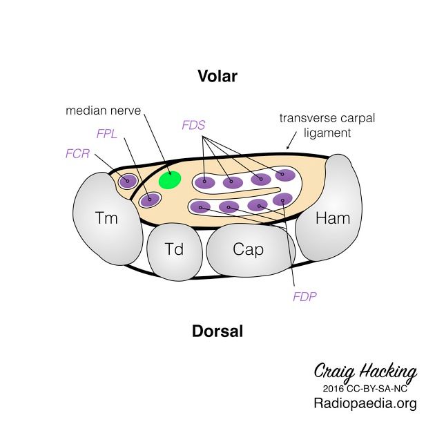
1. The flexor surface of the carpus is deeply concave and is converted into a tunnel by the flexor retinaculum.
2. The 4 tendons of the FDP lie deep, whereas the 4 tendons of the FDS lie superficial. These share a common synovial sheath.
3. The tendon of the FPL lies within its own synovial sheath.
4. The tendon of the FCR lies within its own fibro-osseous tunnel.
5. The median nerve passes beneath the flexor retinaculum b/w the FDS tendon (medial to n.) and FCR tendon (lateral to n.).

Image: Case courtesy of Dr Craig Hacking, Radiopaedia.org. From the case rID: 47155 [Accessed 20 Apr 2020].
Reference(s)
R.M.H McMinn (1998). Last’s anatomy: regional and applied. Edinburgh: Churchill Livingstone.
Gray, H., Carter, H.V. and Davidson, G. (2017). Gray’s anatomy. London: Arcturus.